
Ultrasound
AKA Sonography.
Ultrasound is a safe, painless, non-invasive procedure that uses sound waves to examine the structure and function of soft tissues. It can be used to diagnose a variety of conditions, monitor blood flow or blockages, analyze glandular nodules, or evaluate the veins for grafting into a coronary bypass. It can be used to evaluate the reproductive system or to view an embryo or fetus within a woman's uterus to assess growth and development.
Ultrasound is invaluable to physicians in determining the risk factor of stroke or embolism by detecting the presence of plaque or blood clots.
Because ultrasound provides real-time images (images that are renewed continuously), it can also be used as a guide through procedures such as needle biopsies or to aid in the placement of a needle or catheter.
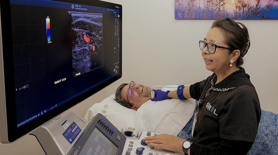
SMIL offers the following Ultrasound scans:
Abdominal Ultrasound - Abdominal Ultrasound imaging shows the organs and other structures in the upper abdomen to help diagnose a variety of conditions, such as abdominal pain or distention, abnormal liver function, enlarged abdominal organs, stones in the gallbladder or kidneys, or an aneurysm in the aorta.
Breast Ultrasound - Breast Ultrasounds are most often used along with a mammogram to examine certain breast changes that can't be clearly seen on a mammogram due to dense tissue in the breasts and other factors.
Carotid Ultrasound - A Carotid Ultrasound uses sound waves to examine the structure and function of the carotid arteries in the neck, which deliver blood from the heart to the brain. Carotid ultrasounds can also be performed to check the state of the carotid artery after surgery, and to verify the position of stents.
Hysterosonogram - A Uterus Ultrasound, or Hysterosonogram, is a valuable technique for examining uterine abnormalities such as polyps, fibroids, endometrial adhesions, or congenital defects.
Obstetric Ultrasound - Obstetric Ultrasounds are useful to establish the presence of a living embryo or fetus, estimate the age of the pregnancy, asses the fetus for normal growth and development, and diagnose any congenital abnormalities of the fetus.
Pelvic Ultrasound - Pelvic Ultrasound imaging shows the organs and other structures in the lower abdomen and pelvis via abdominal (transabdominal) ultrasound; vaginal (transvaginal, endovaginal) ultrasound for women to evaluate the uterus, cervix, and ovaries; and rectal (transrectal) ultrasound for evaluation of the prostate gland in men.
Testicular Ultrasound - Testicular Ultrasounds are used to locate and evaluate masses in the testicles, epididymis, and scrotum; to diagnose causes of testicular pain or swelling; to look for the location of an un-descended testis, or evaluate the cause of infertility such as varicocele.
Thyroid Ultrasound - A Thyroid Ultrasound exam can help diagnose a thyroid condition, analyze the appearance of thyroid nodules, or monitor any changes in thyroid nodules.
Ultrasound Guided Breast Biopsy - An Ultrasound Guided Breast Biopsy uses a fine needle to remove cells from a suspicious area of the breast, which are then examined under a microscope by a pathologist to determine a diagnosis.
Vascular Carotid Ultrasound - A Vascular Carotid Ultrasound can detect blocking or narrowing of the carotid artery in patients with high blood pressure, diabetes, elevated cholesterol, or a family history of stroke or heart disease. It can also be used to check the state of the carotid artery after surgery, and verify the position of stents.
Vascular Ultrasound - Vascular Ultrasounds are used to monitor the blood flow to organs and tissues throughout the body, to locate and identify blockages, abnormalities, or blood clots in the major veins of the legs or arms (deep vein thrombosis), and to determine the source and severity of varicose veins.
Venous Ultrasound - Venous Ultrasounds help Radiologists search for blood clots in the veins of the leg; to determine the cause of long-standing leg swelling which may indicate the presence of varicose veins; and to map out the veins in the leg or arm so that pieces may be used to bypass a narrowed or blocked blood vessel elsewhere.
During an ultrasound, you will be positioned on an examination table that can be tilted or moved. A clear water-based gel is applied to the skin to eliminate air pockets between the transducer and the skin that can block the sound waves from passing into your body. The sonographer (ultrasound SMIL Technologist) or Radiologist then presses the transducer firmly against the skin in various locations, sweeping over the area of interest or angling the sound beam from a farther location to see an area of concern better.
When the examination is complete, you may be asked to dress and wait while the ultrasound images are reviewed. Most ultrasound examinations are completed within 30 minutes to an hour.
You should wear comfortable, loose-fitting clothing for your SMIL ultrasound exam. You may need to remove all clothing and jewelry in the area to be examined, and you may be asked to wear a gown during the procedure. To learn more about your particular Ultrasound exam, select your type below.
For standard diagnostic ultrasound, there are no known harmful effects on humans. Ultrasound exams do not use ionizing radiation.
SMIL locations providing US services:
NORTH SCOTTSDALE
- 92ND ST. & MOUNTAIN VIEW
- 92ND ST. & MOUNTAIN VIEW - Breast Center
- THOMPSON PEAK PKWY & SCOTTSDALE RD
SOUTH SCOTTSDALE
PARADISE VALLEY
PHOENIX
- HIGHLAND & 22ND STREET
- TATUM BLVD & 101
- I-17 & DOVE VALLEY 101 AZ & 31 AVE.
- TATUM & UNION HILLS
- 3RD ST. & DUNLAP
PEORIA
MESA
GILBERT
FOUNTAIN HILLS
SMIL Southwest Medical Imaging Services