Welcome to the Breast Center
Breast scans enable Radiologists to assess and diagnose various conditions such as tumors, abscesses, hematomas or cysts.
SMIL offers breast scan services in Scottsdale, Phoenix, Gilbert, Mesa, Paradise Valley and Fountain Hills, Arizona.
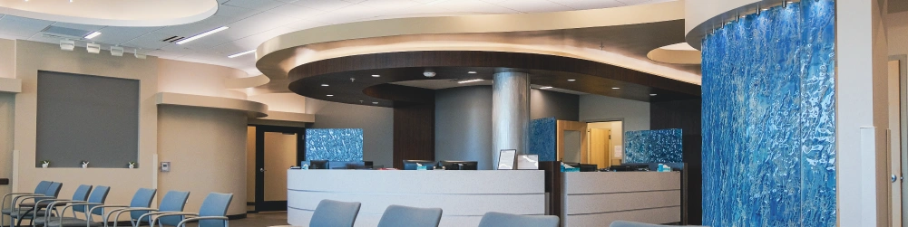
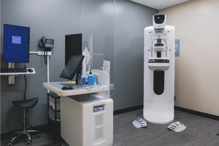
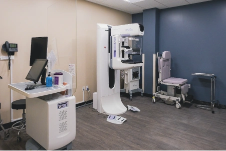

The SMIL Breast Nurse Navigator program, available at all SMIL Breast Centers of Excellence, offers nationally certified, in-person support. Nurse Navigators guide patients through care recommendations from physicians, surgeons, and radiologists providing clarity and compassion throughout their journey.
SMIL offers the following breast scans:
Digital (Screening) Mammography - Digital Mammography is a system that uses low-dose X-rays and electronic detectors (similar to those found in digital cameras) to produce clearer images of the breast than what was previously possible with traditional film mammography, and allows for a more computerized analysis of a breast exam.
MammoScreen® - Mammoscreen® is FDA-cleared, comprehensive breast cancer screening software designed to assist radiologists in reading digital mammograms. It provides support by handling the tedious tasks of reading exams, such as measuring breast density; or by acting as a 'second set of eyes' for the radiologist by highlighting suspicious areas. While AI can never replace the expertise of a radiologist, it is a valuable tool that enhances the confidence and precision of the physicians caring for you. And consistent with our patient-first philosophy, this technology is included at no added cost to patients.
3D Mammograms (Tomosynthesis) - Breast Tomosynthesis (3D Mammography) is used to take 3D images of the breasts, allowing areas of interest to be examined from all angles. This exam is performed at the same time as a 2D mammogram, minimizing time and exposure for the patient. We offer 3D mammography at all of our breast service locations.
Galactography - Galactography, or Ductography, is a highly specialized procedure that uses mammography and a contrast material to obtain images of the inside of the breasts milk ducts.
Needle Localizations - A needle localization of the breast is done when another breast exam shows lumps or abnormalities that require surgical removal. Imaging is used to place a thin wire next to the area of concern in the breast as a guide for the surgical team.
Diagnostic Mammograms - When you have a new breast problem or when extra views of the breast are needed to clarify information from a screening mammogram.
Breast Ultrasound - Often used to examine certain breast changes that can't be clearly seen on a mammogram. Breast ultrasound is most often used along with a mammogram and is not a screening method for breast cancer.
Breast MRI Scans - MRI is helpful in finding abnormalities that are not visible with mammography or ultrasound. In general, MRI is used only in women at high risk for breast cancer.
Breast Biopsies - Breast biopsies are performed to remove a small amount of cells from a suspicious area in the breast for examination in a laboratory. Guided biopsies can be performed in conjunction with mammography (stereotactic), MRI, or ultrasound.
In addition to these services, SMIL offers interventional radiology and nuclear medicine services for breasts:
FAQs
Experiences with mammogram pain vary from person to person. While many describe it as a brief, manageable pressure (similar to a blood pressure cuff), others report sharper pain. Discomfort is highly individual and largely depends on your breast density, breast size, individual pain tolerance, and the skill of the technician.
A large number of patients agree that while the "squish" is awkward, uncomfortable, and occasionally produces a pinching sensation, it is over so fast (about 10 to 30 seconds per image) that they wouldn't classify it as pain. Patients with particularly dense tissue, smaller breasts, or sensitive chests often describe the sensation as intense, pinching, or comparable to a vice.
Tips to Minimize Discomfort
If you are scheduled for your first appointment and are feeling anxious, there are several practical ways to make the experience easier:
Speak up about pain: If the machine pinches, tell your technologist immediately. Many techs will happily readjust or let you control the level of the squeeze.
Time your appointment: Avoid scheduling the screening during the week before your period, as your breasts are naturally more tender and swollen.
Consider skipping caffeine: Some patients find that avoiding coffee and caffeine for a few days leading up to the appointment noticeably reduces breast tenderness.
Ask for modifications: You can ask your technologist if Bella Blankets would be helpful to improve your experience. These are disposable coverlets applied to the mammogram plate that have a slight grip to keep small, dense, or hard-to-position breasts in place, reducing the need for retakes. The soft texture helps prevent skin tears for patients with delicate or thin skin.
Ultimately, while the compression can be unpleasant, most patients agree that the process is incredibly quick and well worth the peace of mind it provides.
You can't wear deodorant, lotion, or powder to a mammogram because they contain metals like aluminum or microscopic particles that create bright white spots on X-ray images. These specks can mimic tiny calcifications, which are early signs of breast cancer. This can lead to a false positive, causing unnecessary anxiety and requiring you to return for additional imaging or biopsies. We do provide wipes in case you accidentally wear deodorant before you arrive, but you may be more comfortable preparing at home.
Medical professionals often use mammograms and ultrasounds together. If a mammogram detects a suspicious area or cannot clearly penetrate a specific area of dense tissue, a follow-up ultrasound is standard.
Mammograms use strictly regulated amounts of X-rays to catch early signs like microcalcifications (stage 0 cancers) that are too small to see with an ultrasound and show up far better on a mammogram. Ultrasounds use sound waves without radiation and are better for evaluating palpable lumps and seeing through dense breast tissue.
The American Cancer Society notes that mammograms are the gold-standard in breast imaging, and the only screening method proven to consistently save lives.
Mammography & Breast Services:
NORTH SCOTTSDALE
- 92ND ST. & MOUNTAIN VIEW
- 92ND ST. & MOUNTAIN VIEW - Breast Center
- THOMPSON PEAK PKWY & SCOTTSDALE RD
SOUTH SCOTTSDALE
PARADISE VALLEY
PHOENIX
PEORIA
MESA
GILBERT
FOUNTAIN HILLS