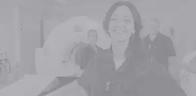
Nuclear Medicine
SCHEDULE NOW
Nuclear medicine imaging provides doctors with information about both structure and function of the region of the body being examined. Nuclear medicine procedures often identify abnormalities early in the progress of a disease and long before medical problems are apparent with other diagnostic tests. Nuclear medicine imaging procedures are noninvasive, with the exception of an intravenous catheter, or IV, are painless and rarely associated with significant discomfort or side effects. Nuclear medicine uses very small amounts of radioactive materials called radiopharmaceuticals or radiotracers to diagnose disease. These radiotracers are injected, inhaled as a gas or ingested and accumulate in the organ or part of the body being examined. Once the tracers are administered, imaging can be started immediately or after a designated period of time using a special imaging device called a gamma scintillation camera that produces pictures for evaluation by the nuclear medicine physician.
Today nuclear medicine offers procedures that are essential in many medical specialties.
SMIL offers nuclear medicine services in Scottsdale, Gilbert, Paradise Valley and Arizona.
SMIL offers the following nuclear medicine services:
Myocardial Perfusion Imaging (MPI)
Myocardial perfusion Imging or MPI is used to diagnose and assess coronary artery disease. It can be used to evaluate unexplained chest pain and other abnormalities of the heart. MPI can also be used to visualize blood flow patterns to the heart, determine the extent of injury to the heart following a heart attack, and evaluate results after heart procedures. This type of imaging is done in two phases: stress and rest. During the resting phase, a radiotracer is injected into the bloodstream through a vein in the arm and images are acquired after a certain amount of time. For the stress phase, the radiotracer is injected after the blood flow into heart muscle is maximized by either physical exercise on a treadmill or by a drug. After a certain amount of time, images are acquired. The nuclear medicine physician and/or cardiologist evaluate the images by comparing the stress and rest blood flow into the heart muscle.
Hepatobiliary Scan
Hepatobiliary imaging allows SMIL nuclear medicine physician to evaluate the the liver, gallbladder, and the ducts that are part of the biliary system. A hepatobiliary scan is also used to diagnose abdominal pain that may be caused by a sudden inflammation of the gallbladder or a poorly functioning gallbladder.
An injection of a radiotracer is given through an IV that is inserted into your arm. After a brief period of time, you will lie on a table and a gamma camera is placed over your abdomen in the area of the liver. This tracer starts out being taken up in the liver by the cells that produce bile and once it reaches the liver, it begins to follow or trace the path of bile. Over time, it completely leaves the liver and shows up in the biliary ducts, gallbladder, and eventually in the small intestines. Images are acquired of this tracer "following" it in the biliary system over a designated period of time.
If your physician wants to evaluate the function of the gallbladder, he or she may ask to have a hepatobiliary scan with ejection fraction or with CCK. When this exam is performed, the same images of the gallbladder are taken. When most of the tracer has left the liver and the gallbladder is filled with this tracer a medicine called cholecystokinin or CCK for short is given slowly over a period of time. Computer calculations of the images taken of the gallbladder over that period of time help the nuclear medicine physician determine the function of the gallbladder. Sometimes, the infusion of CCK may cause slight abdominal discomfort or nausea.
It is important that you inform the nuclear medicine SMIL Technologist of any medications you make be taking as some may affect the results of this exam.
Infection Imaging
Nuclear medicine infection imaging is performed to locate areas of infection in the body. It can help Radiologists locate an infection, determine the origin of the infection, the extent of the infection and assist in determining treatment options. Other than startting an IV into a vein in the arm infection imaging is painless.
A radiotracer is attached or "tagged" to the white blood cells and injected into the bloodstream though the IV. This tagging process is performed at an offsite radiopharmacy and takes several hours. During that time, you may leave the SMIL facility. After the injection, you will be instructed to return at a specific time the next day for imaging. That delay allows the tracer to circulate throughout the body and be taken up in the area of infection. Because of the detailed nature of this type of imaging, the second day you are at SMIL, make take about two hours.
Nuclear Bone Scan
A nuclear bone scan can identify fractures, tumors and diseases of the skeletal system. Its greatest strengths include providing early physiologic information about injury or surgical sites and evaluating large areas - or the entire body - in a single exam. This is particularly well-suited for patients at risk for metastasis to the bone or to monitor therapy for bone metastasis.
PET-CT
By combining images obtained through PET (Positron Emission Tomography) and CT medical imaging is entering a new era. SMIL was the first medical imaging practice in the Phoenix metro area to offer this diagnostic advance. In oncology, PET-CT helps detect tumors at their earliest, most treatable stage. CT provides an anatomical map of the body while PET evaluates the biochemical activity allowing us to see the cells “in action.” Bringing these together the Radiologist is able to accurately identify the tumor and how far it has spread. This makes PET-CT a valuable tool for the clinical evaluation of patients with most cancers, including head and neck cancer, esophageal cancer, lung cancer, breast cancer and colorectal cancer. PET-CT is also used to determine the stage of cancer accurately and to evaluate the effectiveness of cancer therapy. A final oncologic utility of PET-CT is the ability to differentiate benign from malignant tumors.
A break-through application of PET-CT is in neuroradiology, where the technology is used in evaluating patients with cognitive impairment. It can help identify between different dementias and other causes of cognitive decline.
Multi Gated Acquisition Scan (MUGA)
A MUGA scan is a nuclear medicine test designed to evaluate the function of the right and left ventricles of the heart. When a MUGA scan is performed, a small amount of a radioactive isotope is attached to red blood cells and then injected into the blood stream. The isotope emits weak gamma rays which can be detected with a special camera positioned over the patient.
Electrodes are placed in three locations on the chest to detect the electrical activity of the heart and that electrical activity triggers the camera to detect those weak gamma rays.
For approximately 10 minutes multiple images are acquired during each heartbeat and with computer analysis, this ejection fraction determines how effective the heart is pumping the blood out to the rest of the body. There are a number of reasons for such a scan to be ordered, ranging from evaluating heart function during chemotherapy treatments to a thorough examination of a patient who has recently experienced a serious cardiac event.
Renal Scintigraphy
Renal scintigraphy is a form of imaging using radiopharmaceutical or radio tracers that evaluate the function of the kidneys.
SMIL performs multiple renal scintigraphy scans and each is based on a specific diagnosis.
-
Renal perfusion and function imaging examines the blood flow to the kidneys through images taken over 30 minutes
-
Diuretic Imaging detects kidney blockages through images taken before and after the introduction of a diuretic, to move urine through the kidneys
-
ACE-inhibitor renal imaging helps to determine if the cause of a patient’s high blood pressure is coming from the kidneys by comparing kidney images before and after receiving an injection of a blood pressure medicine called an “ACE-inhibitor.”
-
Cortical renal imaging detects the amount of functioning kidney tissue through images taken with a gamma camera approximately three hours after the injection of a radiotracer. At SMIL this scan is done using a SPECT-CT camera located at our Paradise Valley and Scottsdale locations.
SPECT-CT
Single Phonton Emission Computed Tomography (SPECT) uses a gamma camera to create images of the body in three different planes and helps the nuclear medicine physician to "see around" those body parts that may be in the way. The gamma camera rotates around the body collecting data during each stop of the rotation. A computer takes all of that data and creates images, or slices, of the body looking front to back, side to side and top to bottom. By combining the functionality of SPECT imaging and the anatomical detail of a CT scan, the nuclear medicine physician is able to precisely
pinpoint the exact location of that “hot-spot” in an organ or bone.
Thyroid Uptake and Scan
The thyroid gland, located in the neck, uses iodine for the production of thyroid hormones essential to the normal function of the body. By using a small amount of a radioactive form of iodine that emits unseen gamma rays, a special camera called a gamma camera is able to the see size and function of the thyroid gland. Sometimes, nodules may grow on the thyroid gland and this exam will demonstrate whether those nodules are "hot" or "cold"; meaning whether they absorb the iodine (hot) or not (cold). The iodine in a capsule form is swallowed with a small amount of water and is absorbed into the thyroid gland. You will be asked to return at six and 24 hours after that capsule was given to you so that measurements can be made. For the measurements taken at six hours, the gamma camera is placed in three different positions over the neck and one position on the thigh to measure how much iodine is still circulating in the body outside of the thyroid gland. About 24 hours after you took the capsule one measurement is taken over the neck and one over the thigh. Each of the measurements will take three minutes.